
Specialised Women’s Imaging
At Fraser Coast Radiology we offer:
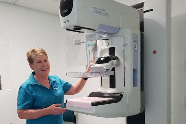
3D Mammography
Mammography is an examination of the breast tissue to detect and diagnose breast pathology. At Fraser Coast Radiology we offer 3D tomosynthesis which is an advanced mammography system allowing low dose imaging with superior image quality.
Early detection of breast cancer
Doctors and scientists agree that early detection is the best defence against breast cancer. Successful treatment and survival rates for patients are dramatically affected by early detection. If lesions are found early, before spreading to lymph nodes, the five-year survival rate is almost 100 percent.
Until now, the best way to do that has been with digital mammography. Digital mammography provides a 2-dimensional picture of the breast and is still one of the most advanced technologies available today.
However, the breast is a 3-dimensional object composed of different structures, such as blood vessels, milk ducts, fat, and ligaments. All of these structures, which are located at different heights within the breast, can overlap and cause confusion when viewed as a 2-dimensional, flat image. Overlapping tissue may be a reason why small breast lesions may be missed or normal tissue may appear abnormal, leading to unnecessary anxiety for patients if called back for further testing.
What is 3D mammography?
3D mammography is a new technology in the fight against breast cancer that allows doctors to examine your breast tissue one layer at a time. 3D mammography uses high-powered computing to convert digital breast images into a stack of very thin layers or “slices”– building what is essentially a “3-dimensional mammogram”. A good analogy for 3D mammography is like thinking of the pages in a book. If you look down at the cover you cannot see all of the pages – but when you open it up, you can go through the entire book page-by-page to see everything between the covers. 3D mammography is designed with the same concept in mind.
Very low X-ray energy is used during the examination so your radiation exposure is below the guidelines. Using 3D mammography and 2D digital mammography together has been proven to significantly reduce “call-backs” by 20-40%. In addition, 3D mammography finds cancers earlier than 2D mammography alone, with a 27% increase in cancer detection and a 40% increase in invasive cancer detection.
What to expect during your exam
A 3D mammography exam is very similar to a traditional mammogram. Just as with a 2D digital mammogram, the technologist will position you, compress your breast under a paddle and take images from different angles. At Fraser Coast Radiology we use soft touch pads to reduce discomfort of compression of your breast.
During the 3D mammography part of the exam, the X-ray arm sweeps in a slight arc over the breast, taking multiple breast images in just seconds. Your doctor is then able to view your breast tissue in one millimetre layers. Instead of viewing all the complexities of your breast tissue in one flat image, the doctor can examine the tissue one page or “layer” at a time.
There is no additional compression required with 3D mammography, and it only takes a few seconds longer for each view. The technologist will view the images at their computer workstation to ensure they have captured adequate images for review by a radiologist, who studies them and reports results to your doctor.
How do I prepare for my Mammogram?
On the day of your appointment please ensure that you do not use Spray-on Deodorant/Perfume or Talcum Powder.
Please arrive 10 minutes early to complete the Mammography Questionnaire.
What to tell us?
Please advise us if you are pregnant, suspect you may be pregnant, are breastfeeding or have breast implants. If your doctor has referred you for a breast ultrasound please also advise us of this when making your appointment.
If you have had previous breast studies please bring any films, reports, or computer disks from these with you to your appointment.
How long does it take?
Depending on the study requested by your doctor your scan will take between 20 and 30 minutes to complete.
What will it cost?
If you have a current Medicare card and referral form from your health practitioner your scan may be Bulk-billed. Medicare rebates depend on the type of study requested by your doctor. If your scan has an out-of-pocket expense, we will let you know when you book your appointment.
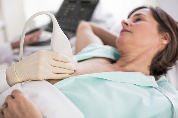
Breast Ultrasound
Ultrasound imaging of breast tissue uses sound waves to produce pictures of the internal structures of the breast. Ultrasound is a safe, non-invasive procedure and does not use ionising radiation.
Your doctor will sometimes refer you for a breast ultrasound to be performed in conjunction with a Mammogram.
How long does it take?
Depending on a number of factors your scan will take between 20 and 30 minutes to complete.
What will it cost?
If you have a current Medicare card and referral form from your health practitioner your scan may be Bulk-billed. Medicare rebates depend on the type of study requested by your doctor. If your scan has an out-of-pocket expense, we will let you know when you book your appointment.
Breast MRI
Breast MRI is recommended for women who are at a high risk for developing breast cancer, usually due to a strong family history or gene mutation such as BRCA1 or BRCA2. It can also assist diagnosis in women with dense breast tissue. Breast MRI is considered more sensitive in detecting breast cancer, and is best performed in combination with mammography and ultrasound.
How long does it take?
Normally your scan will take between 40 minutes to complete.
What will it cost?
Medicare will only pay a rebate under very strict conditions. If your scan meets these conditions, we will let you know when you book your appointment. Otherwise we will inform you of your out-of-pocket expenses at that time.
Interventional Breast Procedures
At Fraser Coast Radiology we offer the following interventional breast procedures:
- Fine Needle Aspiration (FNA)
- Core Biopsy
- Hook wire insertion
- Breast Aspiration